Imaging Tests for Liver Cancer Diagnosis

Accurate imaging techniques play an important role in facilitating liver cancer diagnosis. Source: Shutterstock.
Like most other cancers, early detection is crucial for the successful treatment and recovery of liver cancer. Accurate imaging is important for diagnosing, staging and planning treatment strategies for primary liver cancer. This article highlights the different types of imaging scans used for liver cancer diagnosis.
Imaging scans are non-invasive diagnostic tools that help:
- Visualize chronic liver diseases, such as hepatitis and cirrhosis
- Identify abnormal growths in the liver, including hepatocellular carcinoma (HCC), cholangiocarcinoma and secondary liver cancer
- Distinguish cancerous from non-cancerous growths
- Guide needle aspiration for biopsy
- Assess liver damage
- Guide treatment decisions, such as evaluating eligibility for liver transplantation
- Determine the extent of malignancy and staging
What is the best imaging test for liver cancer?
Currently, there is no standard guideline for diagnosing different types of liver cancer. Selecting the best imaging test for liver cancer diagnosis depends on factors such as:
- Ability to visualize small or hard-to-reach tumors and detect cancer spread beyond the liver
- Availability of scanners and trained professionals for accurate result interpretation
- The risks of each imaging technique, such as radiation exposure from CT scans and potential allergic reactions or kidney complications from contrast agents in some individuals
- Cost consideration and insurance coverage
Liver ultrasound
Abdominal ultrasound is often the first-line imaging test due to its affordability and wide availability. It uses sound waves to create live images of the liver and detect liver tumors. However, it is less sensitive in detecting small tumors, particularly in the early stages of liver cancer.
For suspicious tumors, whether they might be HCC, other types of liver cancer or metastatic liver cancer that cannot be conclusively diagnosed by an ultrasound scan, patients are referred for CT, MRI, or contrast-enhanced ultrasound (CEUS).
Liver CT scan
Computed tomography (CT) scans provide detailed cross-sectional images, which help assess tumor size, location and spread. It is effective in staging liver cancer but exposes patients to radiation.
Liver MRI
Magnetic Resonance Imaging (MRI) offers better contrast for soft tissues, making it easier to distinguish cancerous and non-cancerous lesions. In cases of fatty liver, it helps differentiate tumors from surrounding tissues. Both CT and MRI are more expensive, so they are typically reserved as problem-solving tools for difficult-to-diagnose cases.
Contrast-enhanced ultrasound
The use of contrast-enhanced ultrasound (CEUS) allows visualization of blood flow patterns in the liver. Although there are rare contraindications of allergies to the contrast dye, CEUS is generally well-tolerated. Contrast dye can also be used with CT and MRI, especially for liver cancer staging
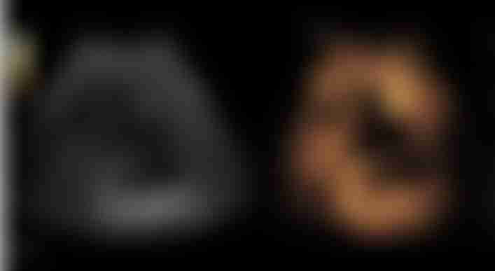
What does liver cancer look like on ultrasound? Left: 2D ultrasound without contrast; right: contrast-enhanced ultrasound liver abscess. Source: Wikimedia Commons.
Other imaging techniques for liver cancer diagnosis
Examples of other imaging techniques are positron emission tomography (PET) and angiography. They are often used in the diagnosis of more complex liver cancers.
PET scans detect cancer spread (metastasis), whereas angiography helps doctors visualize the blood vessels around the liver. It shows how much blood goes to the tumors and helps with planning surgery or liver transplants.
While imaging tests provide critical information in cancer detection, a definitive diagnosis of liver cancer often requires a biopsy. This procedure involves the extraction of tissue samples from the liver for microscopic examination.
How is liver cancer diagnosed early?
Liver cancer surveillance is vital for people at high risk. The six-monthly abdominal ultrasound serves as the primary screening tool. Combining liver ultrasound with alpha-fetoprotein (AFP) blood tests improves early detection.
Learn more: Liver Cancer Screening for High-Risk Individuals
FAQs about imaging tests for liver cancer diagnosis
1. Can an ultrasound detect early liver cancer?
Detecting liver cancer early is usually challenging because symptoms do not appear until the later stages. Additionally, small tumors are difficult to detect during physical exams as the liver is largely covered by the rib cage. However, a review study involving over 6,500 adults found that CEUS can accurately detect liver tumors of any size more than 70% of the time.
2. Is an ultrasound sufficient for diagnosing liver cancer?
An ultrasound is usually the first-line test that picks up suspicious lesions that may be liver cancer. However, it is not always enough by itself for doctors to make the final diagnosis. The ultrasound results depend on the skill of the person operating it. It may also be less accurate in individuals with higher abdominal/visceral fat. By combining contrast dye and other indication tests like the alpha-fetoprotein (AFP) blood test, the detection of early-stage liver cancer can be improved by over 60%.
3. Is CT or MRI better for diagnosing liver cancer?
Both CT and MRI are similar with regard to how well they can detect liver tumors. Doctors consider the use of these tests depends on factors and reasons mentioned before. Usually, a referral for CT or MRI occurs after an ultrasound shows something suspicious.
4. Can a fatty liver look like cancer on a CT scan?
A fatty liver may resemble certain types of liver tumors on a CT scan. In such cases, further evaluation, such as MRI or contrast-enhanced imaging, is necessary to distinguish a fatty liver from cancer accurately.